Alise Rugh
  • Blog
  • About
  • Contact

Fallen Arches Causes And Symptoms

3/31/2015

 
Overview

Adult Acquired Flat Feet

Pes planus can be part of normal development. Infants typically have a minimal arch. Many toddlers have flattening of the long arch, with forefoot pronation and heel valgus on weight-bearing. There may be ligamentous laxity, which is probably determined genetically. Most of these children spontaneously develop a strong normal arch by around age 10.


Causes

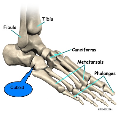
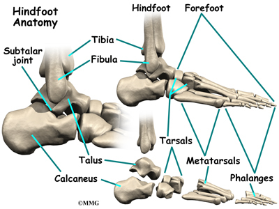
Flat feet in adults can arise from a variety of causes. Here are the most common. An abnormality that is present from birth, stretched or torn tendons, damage or inflammation of the posterior tibial tendon (PTT), which connects from your lower leg, along your ankle, to the middle of the arch, broken or dislocated bones. Some health conditions, such as rheumatoid arthritis, Nerve problems. Other factors that can increase your risk include obesity, diabetes, ageing and Pregnancy.


Symptoms

The primary symptom of fallen arches is painful or achy feet in the area in which the foot arches or on the heel. This area may become swollen and painful to stand still on. This causes the patient to improperly balance on their feet which in turn will cause other biomechanical injuries such as back, leg and knee pain.


Diagnosis

Your doctor examines your feet to determine two things, whether you have flat feet and the cause or causes. An exam may include the following steps, Checking your health history for evidence of illnesses or injuries that could be linked to flat feet or fallen arches, Looking at the soles of your shoes for unusual wear patterns, Observing the feet and legs as you stand and do simple movements, such as raising up on your toes, Testing the strength of muscles and tendons, including other tendons in the feet and legs, such as the Achilles tendon or the posterior tibial tendon, Taking X-rays or an MRI of your feet.


Non Surgical Treatment

Some patients with flat feet may automatically align their limbs in such a way that unpleasant symptoms never develop. In such cases treatment is not usually required. Pain in the foot that is caused by flat feet may be alleviated if the patient wears supportive well-fitted shoes. Some patients say that symptoms improve with extra-wide fitting shoes. Fitted insoles or orthotics (custom-designed arch supports) may relieve pressure from the arch and reduce pain if the patient's feet roll or over-pronate. The benefits of an orthotic only exist while it is being worn. Patients with tendonitis of the posterior tibial tendon may benefit if a wedge is inserted along the inside edge of the orthotic - this takes some of the load off the tendon tissue. Wearing an ankle brace may help patients with posterior tibial tendinitis, until the inflammation comes down. Rest, doctors may advise some patients to rest and avoid activities which may make the foot (feet) feel worse, until the foot (feet) feels better. A combination of an insole and some kind of painkiller may help patients with a ruptured tendon, as well as those with arthritis. Patients with a ruptured tendon or arthritis who find insoles with painkillers ineffective may require surgical intervention. Patients, usually children, whose bones did not or are not developing properly, resulting in flat feet from birth, may require surgical intervention to separate fused bones (rare). Bodyweight management, if the patient is obese the doctor may advise him/her to lose weight. A significant number of obese patients with flat feet who successfully lose weight experience considerable improvement of symptoms.
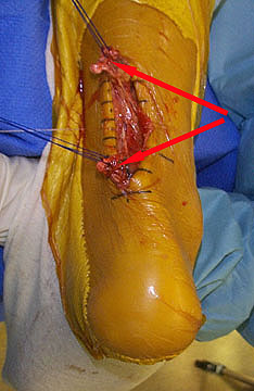


Surgical Treatment

Adult Acquired Flat Foot

This is rare and usually only offered if patients have significant abnormalities in their bones or muscles. Treatments include joint fusion, reshaping the bones in the foot, and occasionally moving around tendons in the foot to help balance out the stresses (called tendon transfer). Flat feet and fallen arches are common conditions that are in most cases asymptomatic. However, in patients who do have symptoms, treatments are available that can help reduce pain and promote efficient movement. Orthotic devices are well recognised as an excellent treatment and podiatrists can offer these different treatment modalities as individualised treatments for patients.


Prevention

Going barefoot, particularly over terrain such as a beach where muscles are given a good workout, is good for all but the most extremely flatfooted, or those with certain related conditions such as plantar fasciitis. Ligament laxity is also among the factors known to be associated with flat feet. One medical study in India with a large sample size of children who had grown up wearing shoes and others going barefoot found that the longitudinal arches of the bare footers were generally strongest and highest as a group, and that flat feet were less common in children who had grown up wearing sandals or slippers than among those who had worn closed-toe shoes. Focusing on the influence of footwear on the prevalence of pes planus, the cross-sectional study performed on children noted that wearing shoes throughout early childhood can be detrimental to the development of a normal or a high medial longitudinal arch. The vulnerability for flat foot among shoe-wearing children increases if the child has an associated ligament laxity condition. The results of the study suggest that children be encouraged to play barefooted on various surfaces of terrain and that slippers and sandals are less harmful compared to closed-toe shoes. It appeared that closed-toe shoes greatly inhibited the development of the arch of the foot more so than slippers or sandals. This conclusion may be a result of the notion that intrinsic muscle activity of the arch is required to prevent slippers and sandals from falling off the child?s foot.


After Care

Time off work depends on the type of work as well as the surgical procedures performed. . A patient will be required to be non-weight bearing in a cast or splint and use crutches for four to twelve weeks. Usually a patient can return to work in one to two weeks if they are able to work while seated. If a person's job requires standing and walking, return to work may take several weeks. Complete recovery may take six months to a full year. Complications can occur as with all surgeries, but are minimized by strictly following your surgeon's post-operative instructions. The main complications include infection, bone that is slow to heal or does not heal, progression or reoccurrence of deformity, a stiff foot, and the need for further surgery. Many of the above complications can be avoided by only putting weight on the operative foot when allowed by your surgeon.

Pain In The Arch What Are The Reasons ?

3/28/2015

 
Overview

Flat feet are a common condition. In infants and toddlers, the longitudinal arch is not developed and flat feet are normal. Most feet are flexible and an arch appears when the person stands on his or her toes. The arch develops in childhood, and by adulthood most people have developed normal arches. Most flat feet usually do not cause pain or other problems. Flat feet may be associated with pronation, a leaning inward of the ankle bones toward the center line. Shoes of children who pronate, when placed side by side, will lean toward each other (after they have been worn long enough for the foot position to remodel their shape). Foot pain, ankle pain or lower leg pain, especially in children, may be a result of flat feet and should be evaluated.

Arch Pain

Causes

In most cases, plantar fasciitis develops without a specific, identifiable reason. There are, however, many factors that can make you more prone to the condition. Tighter calf muscles that make it difficult to flex your foot and bring your toes up toward your shin. Obesity. Very high arch. Repetitive impact activity (running/sports). New or increased activity.Although many people with plantar fasciitis have heel spurs, spurs are not the cause of plantar fasciitis pain. One out of 10 people has heel spurs, but only 1 out of 20 people (5%) with heel spurs has foot pain. Because the spur is not the cause of plantar fasciitis, the pain can be treated without removing the spur.

Symptoms

Experiencing chronic pain of any kind can lead to feelings of fatigue, irritability and even depression. Friends may joke about having 'tired dogs' after a long day, but this is completely different from your experience if arch pain has begun to impact your life on a daily basis. You may dread getting out of bed in the morning and wonder how you're going to get through a work day without having to limp home at the end of it.

Diagnosis

The doctor will examine your feet for foot flexibility and range of motion and feel for any tenderness or bony abnormalities. Depending on the results of this physical examination, foot X-rays may be recommended. X-rays are always performed in a young child with rigid flatfeet and in an adult with acquired flatfeet due to trauma.

Non Surgical Treatment

In mild cases of flatfoot the first line of treatment is often custom orthotics. In patients with a flexible deformity, supporting the arch with a custom arch support will take the strain off the joints and muscles, bringing the heel into a corrected position. Wider shoe gear may be prescribed to accommodate foot pain and motion and stretching exercises to decrease stiffness and stress on the foot. In cases of severe collapse, especially if the patient is not a good surgical candidate or has a mild tear, a brace may be made to accommodate the foot and ankle, thus supporting the arch and ankle.

Arch Pain

Surgical Treatment

Surgery is considered only after 12 months of aggressive nonsurgical treatment. Gastrocnemius recession. This is a surgical lengthening of the calf (gastrocnemius) muscles. Because tight calf muscles place increased stress on the plantar fascia, this procedure is useful for patients who still have difficulty flexing their feet, despite a year of calf stretches. In gastrocnemius recession, one of the two muscles that make up the calf is lengthened to increase the motion of the ankle. The procedure can be performed with a traditional, open incision or with a smaller incision and an endoscope, an instrument that contains a small camera. Your doctor will discuss the procedure that best meets your needs. Complication rates for gastrocnemius recession are low, but can include nerve damage. Plantar fascia release. If you have a normal range of ankle motion and continued heel pain, your doctor may recommend a partial release procedure. During surgery, the plantar fascia ligament is partially cut to relieve tension in the tissue. If you have a large bone spur, it will be removed, as well. Although the surgery can be performed endoscopically, it is more difficult than with an open incision. In addition, endoscopy has a higher risk of nerve damage. The most common complications of release surgery include incomplete relief of pain and nerve damage. Most patients have good results from surgery. However, because surgery can result in chronic pain and dissatisfaction, it is recommended only after all nonsurgical measures have been exhausted.

Prevention

It is possible to prevent arch pain by wearing well-fitting shoes while performing any physical activity. Many times doctors will suggest a therapeutic shoe with a higher heel to relieve the pressure on the achilles tendon and also the arch muscle (plantar fasciitis). People with arch pain suffer from regular flare-ups of pain. However there is no risk to others as this is not a contagious condition.

Stretching Exercises

Ankle evert or strengthening. Lie on your side with your feet hanging off the end of your bed or a weight bench. Bend the toes of the foot that is closer to the ceiling slightly toward your head. This is the starting position. Now raise your toes toward the ceiling while keeping the rest of your leg stationary. Return to the starting position. Reps. 10-15. Now point your toes slightly away from your head. This is the starting position. Raise your toes toward the ceiling. Return to the starting position. Reps. 10-15. Ankle invertor strengthening. Same as above, but do the exercises with the foot that is closer to the floor. Dorsiflexor strengthening. Sit on a desk, table, or counter so that your feet don?t touch the ground. Let your feet dangle comfortably. Bend your foot upward as far as you can comfortably go. Do not let your foot pull inward or outward. Return to the starting position. Reps. 10-15.

Dealing With Pain In The Foot's Arch

3/28/2015

 
Overview

Foot arch pain, or pain on the bottom of the foot can be caused by a number of problems. The arches of the foot control how the forces associated with activities like walking are transferred up and down the leg. If there is a problem with the foot arches or surrounding soft tissues, pain can be felt anywhere from the foot, to the knee, to the lower back. Arch foot pain is also common when standing or walking for long periods. This is often due to weakness and tightness is the muscles and tendons which support the foot arches.

Arch Pain

Causes

The most common cause of arch pain is plantar fasciitis. Plantar fasciitis is the name that describes inflammation of the fibrous band of tissue that connects the heel to the toes. Symptoms of plantar fasciitis include pain early in the morning and pain with long walks or prolonged standing. Arch pain early in the morning is due to the plantar fascia becoming contracted and tight as you sleep through the night. When awakening and walking in the morning, the fascia is still tight and prone to irritation when stretched. When walking or standing for long periods, the plantar fascia becomes inflamed and painful. Treatment of plantar fasciitis is best accomplished with some simple stretching exercises, anti-inflammatory medications, and inserts for your shoes.

Symptoms

The primary symptom is pain or aching in the arch area. This can be accompanied by inflammation and tenderness. If the pain is caused by the plantar fascia, it is likely to be considerably more severe in the mornings due to the muscles being unused. If you notice that the twinges of pain you have are most commonly associated with or immediately after exercise, you might want to visit a good athletic shoe store to make sure you are wearing the right kind of shoe. Another cause of arch pain is plantar fasciitis. The plantar fascia is a tough tissue structure that holds the bottom part of the arch in place. The fascia often becomes inflamed and sore, usually as a result of repetitive motion (for example, very common in those who stand on their feet for work). The pain is often noticeable first thing in the morning and worse with activity.

Diagnosis

The diagnosis of high arch (cavus) foot deformity or Charcot Marie Tooth disease can be made by an orthopedic surgeon in the office. Evaluation includes a thorough history and physical examination as well as imaging studies such as X-rays. The orthopedic surgeon will look at the overall shape, flexibility, and strength of a patient?s foot and ankle to help determine the best treatment. Nerve tests may occasionally need to be performed to help confirm the diagnosis.

Non Surgical Treatment

This is a common foot condition that can be easily treated. If you suffer from arch pain avoid high-heeled shoes whenever possible. Try to choose footwear with a reasonable heel, soft leather uppers, shock absorbing soles and removable foot insoles. When the arch pain is pronation related (flat feet), an orthotic designed with a medial heel post and proper arch support is recommended for treating the pain. This type of orthotic will control over-pronation, support the arch and provide the necessary relief. If the problem persists, consult your foot doctor.

Foot Arch Pain

Surgical Treatment

Patients with adult acquired flatfoot are advised to discuss thoroughly the benefits vs. risks of all surgical options. Most procedures have long-term recovery mandating that the correct procedure be utilized to give the best long-term benefit. Most flatfoot surgical procedures require six to twelve weeks of cast immobilization. Joint fusion procedures require eight weeks of non-weightbearing on the operated foot, meaning you will be on crutches for two months. The bottom line is: Make sure all of your non-surgical options have been covered before considering surgery. Your primary goals with any treatment are to eliminate pain and improve mobility. In many cases, with the properly designed foot orthosis or ankle brace, these goals can be achieved without surgical intervention.

Prevention

Warm up properly. This means not only stretching prior to a given athletic event, but a gradual rather than sudden increase in volume and intensity over the course of the training season. A frequent cause of plantar fasciitis is a sudden increase of activity without suitable preparation. Avoid activities that cause pain. Running on steep terrain, excessively hard or soft ground, etc can cause unnatural biomechanical strain to the foot, resulting in pain. This is generally a sign of stress leading to injury and should be curtailed or discontinued. Shoes, arch support. Athletic demands placed on the feet, particularly during running events, are extreme. Injury results when supportive structures in the foot have been taxed beyond their recovery capacity. Full support of the feet in well-fitting footwear reduces the likelihood of injury. Rest and rehabilitation. Probably the most important curative therapy for cases of plantar fasciitis is thorough rest. The injured athlete must be prepared to wait out the necessary healing phase, avoiding temptation to return prematurely to athletic activity.

Which Are Usually The Principal Brings About Involving Adult Aquired FlatFoot ?

3/27/2015

 
Overview

PTTD is truly a typical situation treated simply by foot along with ankle specialists. Although there will be a role for surgical therapy of PTTD, conservative care often can stop or perhaps delay surgical intervention. Decreasing inflammation as well as stabilizing the affected joints associated with almost all the posterior tibial tendon can decrease pain as well as improve practical levels. Along With many distinct modalities available, aggressive nonoperative strategies needs for you to be considered in the treatment method of PTTD, such as early immobilization, the employment of long-term bracing, physical therapy, as well as anti-inflammatory medications. In your event that these techniques fail, proper evaluation along with work-up for surgical intervention should be employed.

Acquired Flat Feet

Causes

There really are a quantity of theories as to why the particular tendon gets to always be able to be inflamed and also stops working. That could become associated for the poor blood provide inside the tendon. Escalating age, inflammatory arthritis, diabetes along with obesity have been found being causes.

Symptoms

Pain and also swelling around the inside aspect of the ankle initially. Later, the arch of the foot may fall (foot gets flat), this change contributes to walking for you to become challenging and painful, as well as standing for as well long periods. Since the actual flat foot gets established, pain may progress towards the outer a portion of your ankle. Eventually, arthritis may develop.

Diagnosis

Observe forefoot to hindfoot alignment. Perform this with the affected person sitting and the heel throughout neutral, as well as with most the individual standing. I such as to set blocks under the particular forefoot with the heel inside neutral for you to observe how much forefoot correction can be required to aid hold the hindfoot position. 1 last note will be to check just about all joints for stiffness. in cases regarding prolonged PTTD or coalition, rigid deformity can be current then one must carefully examine the actual joints of the midfoot and hindfoot for stiffness as well as arthritis within the surgical pre-planning.

Non surgical Treatment

Nonoperative therapy of stage one and two acquired adult flatfoot deformity may be successful. General components in the remedy include the use involving comfort along with ease shoes. activity modification to become able to stay away from exacerbating activities. weight loss if indicated. Particular components involving treatment that will with time can lead to marked improvement in signs include a higher repetition, low resistance strengthening program. appropriate bracing or a medial longitudinal arch support. When the particular posterior tibial tendon will be intact, a new number of workouts directed at strengthening the actual elongated as well as dysfunctional tendon complex may be successful. in stage 2 deformities, this is mixed having an ankle brace to get a time period involving 2-3 several weeks until the signs resolve. From this point, the affected person is transitioned for an orthotic insert which may assist to support the arch. Inside patients together with stage one deformity it may be achievable to help to make use of an arch assistance immediately.

Acquired Flat Feet

Surgical Treatment

Good in order to excellent results for greater than 80% involving patients happen to become able to be reported with 5 years' followup for the surgical interventions recommended below. However, the postoperative recovery can always be a lengthy process, and most surgical methods need patients to use a new plaster cast with regard to two to three months. Though many patients statement which their particular perform can be well improved through 6 months, within our experience annually is necessary to recuperate genuinely and gain total practical improvement after the surgery. Clearly, several patients aren't candidates with regard to such major reconstructive surgery.

The Causes Along With Treatment

3/25/2015

 
Overview

Most flat feet are not painful, specifically individuals flat feet seen in children. Throughout your adult acquired flatfoot, pain occurs since soft tissues (tendons and also ligaments) have been torn. The Particular deformity progresses or even worsens since when your essential ligaments as well as posterior tibial tendon are lost, nothing may take their own place to carry up the particular arch of the foot. Your painful, progressive adult acquired flatfoot affects females 4x as frequently as men. That occur in middle to older grow older individuals with a mean age of 60 years. The Majority Of individuals who develop the particular condition curently have flat feet. The change occurs in a foot in which the arch actually begins to flatten greater than before, together with pain along with swelling creating on the inside in the ankle. Precisely why this event occur in many people (female greater than male) and only in a foot remains poorly understood. Contributing elements enhancing the likelihood of adult acquired flatfoot are diabetes, hypertension, as well as obesity.

Adult Acquired Flat Foot

Causes

There certainly are generally a quantity of theories concerning why the particular tendon gets inflamed and stops working. The idea could end up being associated towards the poor blood provide within the tendon. increasing age, inflammatory arthritis, diabetes and also weight problems happen to be located to become causes.

Symptoms

Not everyone together with adult flatfoot has problems with pain. Individuals which do usually experience it across the ankle or perhaps within the heel. the pain is actually generally a whole lot worse together with activity, similar to strolling or perhaps standing regarding extended periods. Sometimes, if the issue develops coming from arthritis inside the foot, bony spurs over the top and also side of the foot develop and make wearing shoes much more painful. Diabetic patients need to watch pertaining to swelling or perhaps big lumps inside the feet, because they may not discover just about any pain. These People are also with higher risk pertaining to developing significant deformities via his or her flatfoot.

Diagnosis

The diagnosis of tibialis posterior dysfunction can be essentially clinical. However, plain radiographs in the foot along with ankle are generally useful for assessing the actual degree of deformity also to confirm your presence or perhaps deficiency of degenerative changes within the subtalar and also ankle articulations. The Actual radiographs are also useful to exclude various other brings about involving an acquired flatfoot deformity. The Actual nearly all helpful radiographs tend to be bilateral anteroposterior along with lateral radiographs with the foot and a mortise (true anteroposterior) look at your ankle. Almost All radiographs should be carried out with all the patient standing. Inside most cases we have got seen simply no role for magnetic resonance imaging or even ultrasonography, because the diagnosis can always be produced clinically.

Non surgical Treatment

Stage one deformities usually reply for you to conservative or non-surgical therapy like anti-inflammatory medication, casting, practical orthotics or a foot ankle orthosis known as the Richie Brace. In case these modalities tend to be unsuccessful surgery is actually warranted.

Acquired Flat Foot

Surgical Treatment

In cases associated with PTTD that have progressed substantially or perhaps failed to enhance using non-surgical treatment, surgery might become required. for several advanced cases, surgery might end up being the only real option. The foot along with ankle surgeon will figure out the best approach regarding you.

Achilles Tendonitis

3/23/2015

 
Overview
Achilles Tendinitis The Achilles tendon is the largest tendon in the human body. It connects the calf muscle to the heel bone. However, this tendon is also the most common site of rupture or tendonitis, an inflammation of the tendon due to overuse. Achilles tendon rupture is a partial or complete tear of the Achilles tendon. It comes on suddenly, sometimes with a popping sound, and can be debilitating. A full rupture is more severe, but less common, than a partial rupture. A full rupture splits the Achilles tendon so that it no longer connects the calf muscle to the heel: the calf muscle can no longer cause the foot to push off, so normal walking is impossible. If it is a full rupture, then lightly pinching the Achilles tendon with the forefinger and thumb will reveal a gap in the Achilles tendon. Partial and full Achilles tendon ruptures are most likely to occur in sports requiring sudden stretching, such as sprinting and racquet sports. Partial Achilles tendon tears are also common among middle and long distance runners.

Causes
Inflammation/strain of the tendon is usually caused by overuse, for example, frequent jumping in volleyball, netball or basketball. It is often also caused by a sudden increase in certain types of training, such as hill sprinting or track running, particularly when running in spikes. Tendinopathy can also be associated with ageing. Our ability to regenerate damaged tissue decreases as we age and the quality of the tendon deteriorates. However, the better news is that sensible training can actually strengthen all our soft tissue (tendons, ligaments and muscle). Tightness in the calf muscles will demand greater flexibility of the tendon, which inevitably results in overuse and injury. Biomechanically, the tightness can reduce the range of dorsiflexion (toe up position) in the ankle, which increases the amount and duration of pronation. This problem is known as overpronation.* This reduces the ability of the foot to become a rigid lever at push off and places more lateral and linear forces through the tendon. This imbalance can translate into altered rotation of the tibia (shin bone) at the knee joint and, in turn, produce compensatory rotation at the hip joint with subsequent injuries to the shin, knee and hip. Pronation is part of the natural movement of the subtalar joint in the foot. It allows eversion (turning the sole outwards), dorsiflexion and abduction (pointing the toes out to the side). Pronation is a normal part of the gait cycle, when walking and running, and it helps to provide shock absorption in the foot. When pronation is excessive, the foot has a tendency to roll inward more than normally acceptable. One sign of overpronation is greater wear on the inside of your running shoes than on the midsole. Lack of stability around the ankle joint can also be a contributory factor, as recurrent ankle sprains appear to be associated with a high incidence of Achilles tendonopathy. Wearing shoes that dont fit or support the foot properly can be a major contributing cause of Achilles tendon injury.

Symptoms
Symptoms of an Achilles tendon rupture usually directly follow a traumatic event where the foot is forced in an upward position at the ankle, causing a sudden tight stretch of the Achilles tendon. There can also be a direct blow to the tendon causing a rupture. There is typically a popping feeling or even a popping sound described during the occurance of the rupture. Typically there is pain with swelling in the region. Often the patient is unable to put weight on this foot as there is too much pain.

Diagnosis
When Achilles tendon injury is suspected, the entire lower lag is examined for swelling, bruising, and tenderness. If there is a full rupture, a gap in the tendon may be noted. Patients will not be able to stand on the toes if there is a complete Achilles tendon rupture. Several tests can be performed to look for Achilles tendon rupture. One of the most widely used tests is called the Thompson test. The patient is asked to lie down on the stomach and the examiner squeezes the calf area. In normal people, this leads to flexion of the foot. With Achilles tendon injury, this movement is not seen.

Non Surgical Treatment
Non-surgical treatment of Achilles tendon rupture is usually reserved for patients who are relatively sedentary or may be at higher risk for complications with surgical intervention (due to other associated medical problems). This involves a period of immobilization, followed by range of motion and strengthening exercises; unfortunately, it is associated with a higher risk of re-rupture of the tendon, and possibly a less optimal functional outcome. Achilles Tendon

Surgical Treatment
Some surgeons feel an early surgical repair of the tendon is beneficial. The surgical option was long thought to offer a significantly smaller risk of re-rupture compared to traditional non-operative management (5% vs 15%). Of course, surgery imposes higher relative risks of perioperative mortality and morbidity e.g. infection including MRSA, bleeding, deep vein thrombosis, lingering anesthesia effects, etc.

    Archives

    March 2015

    Categories

    All

    RSS Feed


Powered by Create your own unique website with customizable templates.